당신이 넘어지지 않는 건 ‘뇌’가 아니라 ‘허리’ 때문 Walking on ice takes more than brains

From right: Steeve Bourane, Antoine Dalet, Stephanie Koch, Chris Padilla, Cathy Charles,
Graziana Gatto, Tommie Velasquez and Martyn Goulding
Click here for a high-resolution image.
Image: Courtesy of the Salk Institute for Biological Studies
이번 연구에 참여한 솔크생물학연구소 굴딩 교수팀. 맨 오른쪽이 마틴 굴딩 교수다. - 살크연구소 제공
케이콘텐츠 kcontents
가느다란 평행봉 위에 서거나 얼음판을 걸을 때 넘어지지 않는 이유는 대개 뇌가 평형감각을 유지하기 때문이라고 생각한다. 하지만 이런 균형감각은 뇌가 아닌 척추신경 덕분이라는 사실이 새롭게 밝혀졌다. 마틴 굴딩 미국 솔크생물학연구소 교수팀은 척추신경의 ‘RORα’ 뉴런이 미세한 압력정보를 이용해 아슬아슬한 곳에서 균형을 잡고 걸을 수 있도록 하는 데 핵심적인 역할을 한다는 사실을 밝혀내고 ‘셀’ 1월 29일자에 발표했다. 연구팀은 유전적으로 변형시킨 광견병 바이러스를 실험용 쥐에게 감염시키고 쥐가 발로 느낀 감각이 어떤 통로를 통해 전달되는 지를 관찰했다. 광견병 바이러스는 신경세포를 표적으로 하는 바이러스이기 때문에 연구의 재료로 선택됐다. 광견병 바이러스를 관찰한 결과, 발이 느낀 촉감과 압력에 대한 감각이 척추신경의 ‘RORα’ 신경다발에서 종합된다는 사실이 밝혀졌다. 이곳은 뇌의 운동영역과 연결되는 곳으로, 이전까지 발과 뇌를 잇는 징검다리로 여겨졌다. 연구팀은 유전자조작을 통해 RORα 신경다발이 없는 쥐를 만들고 이동과 균형감각이 정상쥐와 차이가 있는지를 살폈다. 그 결과 평지와 같은 곳에서는 움직임이 둔하긴 했지만 여전히 걷고나 멈춰설 수 있었다. 하지만 좁은 철봉 위를 걷게끔 하자 차이가 두드러졌다. RORα가 결손된 쥐들은 그렇지 않은 쥐들에 비해 균형을 잡고 오르는 감각이 눈에 띄게 떨어졌다. 연구팀은 “이번에 발견한 신경다발이 발에서 오는 정보를 종합해 발이 어떻게 움직여야 하는지 명령을 내리는 데 핵심적인 역할을 한다는 사실이 밝혀졌다”고 설명했다. 동아사이언스 이우상 기자 idol@donga.com |

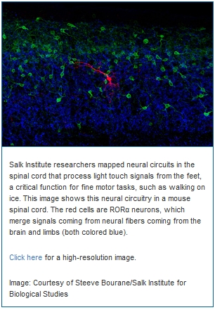
Salk scientists discover how a "mini-brain" in the spinal cord aids in balance LA JOLLA–Walking across an icy parking lot in winter–and remaining upright–takes intense concentration. But a new discovery suggests that much of the balancing act that our bodies perform when faced with such a task happens unconsciously, thanks to a cluster of neurons in our spinal cord that function as a “mini-brain” to integrate sensory information and make the necessary adjustments to our muscles so that we don’t slip and fall. In a paper published January 29, 2015 in the journal Cell, Salk Institute scientists map the neural circuitry of the spinal cord that processes the sense of light touch. This circuit allows the body to reflexively make small adjustments to foot position and balance using light touch sensors in the feet. The study, conducted in mice, provides the first detailed blueprint for a spinal circuit that serves as control center for integrating motor commands from the brain with sensory information from the limbs. A better understanding of these circuits should eventually aid in developing therapies for spinal cord injury and diseases that affect motor skills and balance, as well as the means to prevent falls for the elderly. “When we stand and walk, touch sensors on the soles of our feet detect subtle changes in pressure and movement. These sensors send signals to our spinal cord and then to the brain,” says Martyn Goulding, a Salk professor and senior author on the paper. “Our study opens what was essentially a black box, as up until now we didn’t know how these signals are encoded or processed in the spinal cord. Moreover, it was unclear how this touch information was merged with other sensory information to control movement and posture.” While the brain’s role in cerebral achievements such as philosophy, mathematics and art often take center stage, much of what the nervous system does is to use information gathered from our environment to guide our movements. Walking across that icy parking lot, for instance, engages a number of our senses to prevent us from falling. Our eyes tell us whether we’re on shiny black ice or damp asphalt. Balance sensors in our inner ear keep our heads level with the ground. And sensors in our muscles and joints track the changing positions of our arms and legs. Every millisecond, multiple streams of information, including signals from the light touch transmission pathway that Goulding’s team has identified, flow into the brain. One way the brain handles this data is by preprocessing it in sensory way stations such as the eye or spinal cord. The eye, for instance, has a layer of neurons and light sensors at its back that performs visual calculations–a process known as “encoding”–before the information goes on to the visual centers in the brain. In the case of touch, scientists have long thought that the neurological choreography of movement relies on data-crunching circuits in the spinal cord. But until now, it has been exceedingly difficult to precisely identify the types of neurons involved and chart how they are wired together. In their study, the Salk scientists demystified this fine-tuned, sensory-motor control system. Using cutting-edge imaging techniques that rely on a reengineered rabies virus, they traced nerve fibers that carry signals from the touch sensors in the feet to their connections in the spinal cord. They found that these sensory fibers connect in the spinal cord with a group of neurons known as RORα neurons, named for a specific type of molecular receptor found in the nucleus of these cells. The RORα neurons in turn are connected by neurons in the motor region of brain, suggesting they might serve as a critical link between the brain and the feet. http://www.salk.edu/news/pressrelease_details.php?press_id=2070 |
edited by kcontents
"from past to future"
데일리건설뉴스 construction news
콘페이퍼 conpaper
.


